A cranial nerve examination is a critical component of neurological assessment, evaluating the function of the 12 cranial nerves to identify pathologies affecting sensory, motor, or special functions, requiring detailed anatomical knowledge.
1.1 Importance of Cranial Nerve Examination
The cranial nerve examination is essential for diagnosing neurological conditions, as it provides insights into the brain’s function and structure. Early detection of abnormalities aids in identifying pathologies like nerve palsies, tumors, or infections. It is non-invasive and cost-effective, making it a critical tool in clinical practice. Accurate findings guide further investigations and treatment plans, improving patient outcomes. Additionally, it helps monitor disease progression and response to therapy. This examination is vital for both diagnostic and prognostic purposes, ensuring comprehensive neurological assessment and enhancing clinical decision-making.
1.2 Overview of the 12 Cranial Nerves
The 12 cranial nerves originate from the brain and regulate diverse functions, including sensory perception, motor control, and special functions like taste and balance. Each nerve has distinct roles: the olfactory (smell) and optic (vision) nerves handle sensory input, while the oculomotor, trochlear, and abducens control eye movements. The trigeminal and facial nerves manage facial sensation and expression, and the vestibulocochlear nerve is crucial for hearing and balance. The glossopharyngeal, vagus, and accessory nerves regulate swallowing, heart rate, and neck movements. The hypoglossal nerve governs tongue movements. Together, they are vital for maintaining essential bodily functions and are systematically assessed during a cranial nerve examination.
1.3 Purpose of the Examination
The purpose of a cranial nerve examination is to assess the functional integrity of the 12 cranial nerves, identifying abnormalities that may indicate underlying neurological conditions. This systematic evaluation helps diagnose conditions such as neuropathies, tumors, or traumatic injuries affecting cranial nerve pathways. By testing sensory, motor, and special functions, healthcare providers can localize lesions and determine the extent of nerve involvement. The examination also guides further diagnostic investigations, such as neuroimaging, and informs treatment plans. Accurate findings are essential for improving patient outcomes and managing complex neurological disorders effectively.
Preparation for the Examination
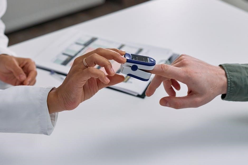
Preparation involves gathering equipment, ensuring patient comfort, and following infection control measures. Introduce yourself, confirm patient identity, and obtain consent. Position the patient appropriately for a thorough assessment.
2.1 Gathering Equipment
Gathering the right equipment is essential for a thorough cranial nerve examination. A torch is needed for assessing pupillary reactions and eye movements. A Snellen chart or near vision card is used to evaluate visual acuity. Non-pungent odor bottles, such as coffee or peppermint, are required for testing the olfactory nerve. A tongue depressor is necessary for eliciting the gag reflex and assessing the glossopharyngeal and vagus nerves. Cotton swabs or a soft brush are used to test facial sensation via the trigeminal nerve. A pen or other object is needed for visual field testing. Ensure all equipment is clean and readily available to maintain efficiency during the examination.
2.2 Patient Positioning and Comfort
Patient positioning and comfort are crucial for an effective cranial nerve examination. The patient should be seated in a comfortable chair, positioned at eye level with the examiner, and approximately 1 meter apart to facilitate accurate visual assessments. Ensure the patient’s arms are relaxed by their sides. A quiet environment is essential for hearing tests. The patient should remove any glasses or contact lenses for visual acuity testing. Proper positioning ensures the patient’s cooperation and minimizes discomfort during the examination. The examiner should also ensure they are at the same level as the patient to accurately assess eye movements and facial expressions. Comfort and clear communication are key to obtaining reliable results.
2.3 Infection Control Measures
Infection control is vital during a cranial nerve examination to protect both the patient and examiner. Handwashing with soap and water or an alcohol-based hand sanitizer is essential before and after the examination. Personal protective equipment, such as gloves, should be worn if there is a risk of exposure to bodily fluids. Instruments like tongue depressors and pens should be used carefully to avoid contamination. Proper disposal of any single-use items is necessary. Maintaining a clean and hygienic environment ensures a safe examination process and prevents the spread of infections. These measures are fundamental to adhering to healthcare standards and patient safety.
Introducing yourself and obtaining consent are crucial steps before performing a cranial nerve examination. Greet the patient politely, state your name, and explain your role. Clearly describe the examination process, ensuring the patient understands its purpose and what to expect. Address any concerns or questions they may have. Obtain explicit consent, respecting their autonomy and right to refuse participation. This step builds trust and ensures a cooperative environment for the examination. Proper communication is key to making the patient feel comfortable and informed throughout the process. It also ensures compliance with ethical and legal standards in healthcare practice.
Examination of Cranial Nerves
The examination systematically evaluates the 12 cranial nerves, assessing sensory, motor, and special functions through specific tests, such as visual acuity, smell identification, and muscle strength assessments.
3.1 Cranial Nerve I: Olfactory Nerve
The olfactory nerve (Cranial Nerve I) is responsible for transmitting sensory information related to smell. To assess its function, the patient is asked to identify non-irritating odors, such as coffee or peppermint, using each nostril separately. This test evaluates the integrity of the olfactory pathway. Normal responses include correct identification of the odor, while abnormal findings may indicate anosmia (loss of smell) or hyposmia (reduced olfactory sensitivity). It is important to avoid using irritating substances, such as ammonia, which can stimulate the trigeminal nerve instead. This nerve is unique as it is the only cranial nerve that bypasses the brainstem, directly connecting to the olfactory cortex.
3.2 Cranial Nerve II: Optic Nerve
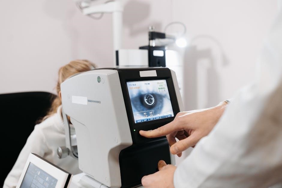
The optic nerve (Cranial Nerve II) is responsible for transmitting visual information from the retina to the brain. Assessment involves evaluating visual acuity using a Snellen chart, examining pupil reactions to light (both direct and consensual), and testing visual fields using confrontation methods. Normal findings include sharp vision, equal pupil reactions, and full visual fields. Abnormalities, such as reduced acuity, afferent pupillary defects, or field deficits, may indicate conditions like optic neuritis or tumors. This nerve is crucial for vision and its integrity is essential for normal visual function. Proper testing ensures early detection of potential neurological or ophthalmological issues.
3.3 Cranial Nerve III: Oculomotor Nerve
The oculomotor nerve (Cranial Nerve III) controls eye movements, pupil constriction, and eyelid elevation. Examination involves assessing eye movements by tracking an object in all directions, testing pupil reactions to light, and evaluating eyelid function. Normal findings include full eye movements, equal pupil reactions, and absence of ptosis. Abnormalities, such as limited eye movement, dilated pupils, or eyelid drooping, may indicate nerve damage, aneurysms, or neurological conditions. Accurate testing ensures early detection of potential issues affecting vision and eye function. This nerve is essential for precise ocular movements and maintaining normal vision, making its examination critical in neurological assessments.
3.4 Cranial Nerve IV: Trochlear Nerve
The trochlear nerve (Cranial Nerve IV) innervates the superior oblique muscle, controlling eye rotation and intorsion. Examination involves assessing eye movements, particularly downward and inward gaze. Patients are asked to look downward while tilting their head to test muscle function. Normal findings include smooth, coordinated eye movements without nystagmus. Abnormalities, such as limited movement, double vision, or head tilt, may indicate nerve damage or conditions like stroke or trauma. Accurate assessment of the trochlear nerve is crucial for identifying issues affecting eye coordination and balance, ensuring proper diagnostic and therapeutic interventions. This nerve plays a key role in maintaining binocular vision and ocular alignment.
3;5 Cranial Nerve V: Trigeminal Nerve
The trigeminal nerve (Cranial Nerve V) is responsible for sensory input from the face and motor control of the muscles of mastication. Examination involves assessing both sensory and motor functions. Sensory testing is done using a cotton swab or blunt object to evaluate light touch, pain, and temperature sensation on the face. Motor function is assessed by observing the patient’s ability to clench their jaw and resist jaw opening. Abnormal findings may include facial numbness, weakness in jaw movements, or altered sensation, which could indicate conditions like trigeminal neuralgia, multiple sclerosis, or stroke. Accurate testing is vital for diagnosing trigeminal nerve dysfunction.
3.6 Cranial Nerve VI: Abducens Nerve
The abducens nerve (Cranial Nerve VI) controls the lateral rectus muscle, enabling eye abduction. Examination involves assessing horizontal eye movements by asking the patient to follow an object with their eyes. Test each eye separately, observing for limitations or nystagmus. Weakness or impairment may result in medial strabismus or inability to abduct the affected eye. Abnormal findings can indicate conditions like intracranial hypertension, stroke, or nerve compression. Accurate testing is essential for diagnosing abducens nerve dysfunction, which may present with diplopia or difficulty moving the eye outward. This nerve is often affected in raised intracranial pressure due to its long intracranial course.
3.7 Cranial Nerve VII: Facial Nerve
The facial nerve (Cranial Nerve VII) governs facial expressions, taste for the anterior two-thirds of the tongue, and the stapedius reflex. Examination involves assessing symmetry in facial movements, such as raising eyebrows, puckering lips, and showing teeth. Test motor strength by resisting the patient’s attempts to close their eyes or smile. Taste testing involves placing sweet, sour, salty, or bitter substances on the anterior tongue. Abnormal findings include facial weakness (e.g., unilateral facial droop), inability to taste, or hyperacusis due to stapedius muscle dysfunction. Lesions can result from conditions like Bell’s palsy, stroke, or tumors affecting the nerve.
3.8 Cranial Nerve VIII: Vestibulocochlear Nerve
The vestibulocochlear nerve (Cranial Nerve VIII) is responsible for hearing and balance. It has two divisions: the cochlear nerve (hearing) and the vestibular nerve (balance). Examination involves assessing hearing with tests like Weber and Rinne tuning fork tests, pure-tone audiometry, or whisper tests. Balance is evaluated using the Dix-Hallpike maneuver to check for vertigo and nystagmus, and the Romberg test to assess equilibrium. Abnormal findings include unilateral hearing loss, tinnitus, or vertigo, suggesting conditions like labyrinthitis, Ménière’s disease, or acoustic neuromas. This nerve’s dysfunction can significantly impact communication and equilibrium, necessitating thorough evaluation and referral for further testing.
3.9 Cranial Nerve IX: Glossopharyngeal Nerve
The glossopharyngeal nerve (Cranial Nerve IX) manages sensory and motor functions, including taste on the posterior one-third of the tongue, pharyngeal sensation, and swallowing. The gag reflex is a key test, where a tongue depressor is used to stimulate the pharynx; absence may indicate nerve dysfunction. Taste can be assessed using non-pungent substances like sucrose or citric acid. Motor function is evaluated by observing the rise of the uvula during a palatal reflex. Lesions may cause difficulty swallowing, reduced taste, or impaired gag reflex, suggesting conditions like strokes, tumors, or infections. Accurate assessment is vital for diagnosing and managing related pathologies.
3.10 Cranial Nerve X: Vagus Nerve
The vagus nerve (Cranial Nerve X) is a mixed nerve with sensory and motor functions, including regulation of visceral organs, taste in the epiglottic region, and motor control of laryngeal and pharyngeal muscles. Examination involves assessing the gag reflex by stimulating the pharynx and observing the rise of the soft palate during speech or yawning. Hoarseness or difficulty swallowing may indicate dysfunction. Lesions can result in vocal cord paralysis or impaired gut motility, often seen in conditions like strokes, tumors, or neuropathies. Accurate evaluation is essential for diagnosing vagal nerve pathologies, which can significantly impact respiratory, digestive, and autonomic functions.
3.11 Cranial Nerve XI: Accessory Nerve
The accessory nerve (Cranial Nerve XI) is a motor nerve responsible for innervating the sternocleidomastoid and trapezius muscles, essential for head rotation, shoulder shrug, and scapular stabilization. Examination involves assessing muscle strength and movement by asking the patient to rotate their head against resistance (testing sternocleidomastoid) and shrug their shoulders (testing trapezius). Muscle atrophy, weakness, or fasciculations may indicate dysfunction. Lesions can result from trauma, neurological disorders, or tumors, leading to impaired mobility and posture. Accurate assessment of CN XI is crucial for diagnosing and managing conditions affecting motor function in the neck and shoulder region.
3.12 Cranial Nerve XII: Hypoglossal Nerve
The hypoglossal nerve (Cranial Nerve XII) controls the motor function of the tongue, enabling speech, chewing, and swallowing. Examination involves assessing tongue protrusion, movement, and strength. The patient is asked to stick out their tongue and move it side-to-side. Atrophy, fasciculations, or deviation from the midline upon protrusion may indicate dysfunction. Lesions can result from neurological conditions, trauma, or tumors, leading to speech and swallowing difficulties. Accurate assessment of CN XII is vital for diagnosing conditions affecting oral motor functions and ensuring proper management of related impairments.
Normal and Abnormal Findings
Normal findings include symmetric responses, full range of motion, and absence of atrophy or fasciculations. Abnormal findings may indicate dysfunction, such as weakness, paralysis, or altered reflexes.
4.1 Normal Responses and Variations
Normal cranial nerve examination findings include symmetric responses, such as coordinated eye movements, normal facial expressions, and intact sensory perception; Variations may occur due to anatomical differences or age-related changes, but these do not indicate dysfunction. For example, mild asymmetry in facial movements or slight differences in olfactory perception are common and benign. Normal findings help rule out cranial nerve pathologies, ensuring accurate diagnosis and appropriate management.
4.2 Signs of Cranial Nerve Dysfunction
Signs of cranial nerve dysfunction vary depending on the affected nerve but may include sensory deficits, motor weakness, or reflex abnormalities. For instance, anosmia (loss of smell) indicates olfactory nerve dysfunction, while visual field defects suggest optic nerve issues. Weakness in eye movements or diplopia may point to oculomotor, trochlear, or abducens nerve impairment. Facial asymmetry or difficulty swallowing could indicate trigeminal, facial, glossopharyngeal, or vagus nerve dysfunction. Hearing loss or balance issues may signify vestibulocochlear nerve damage. Tongue weakness or deviation suggests hypoglossal nerve involvement. These findings guide further investigation and diagnosis of underlying conditions;
Common Pathologies and Clinical Implications
Cranial nerve pathologies include traumatic injuries, infections, neurodegenerative disorders, and tumors, each impacting nerve function and necessitating prompt clinical evaluation for appropriate management and treatment.
5.1 Traumatic Injuries
Traumatic injuries to cranial nerves often result from head trauma, fractures, or penetrating wounds. Commonly affected nerves include the olfactory (CN I), optic (CN II), and facial (CN VII) nerves. Mechanisms may involve direct compression, nerve avulsion, or indirect forces causing nerve stretching or ischemia. Symptoms vary depending on the nerve affected, such as anosmia (CN I), vision loss (CN II), or facial paralysis (CN VII). Diagnosis involves clinical examination and imaging (e.g., MRI or CT scans). Management may include surgical intervention, observation, or rehabilitation. Early recognition and treatment are critical to improving outcomes and minimizing long-term deficits.
5.2 Infections and Inflammatory Conditions
Infections and inflammatory conditions affecting cranial nerves can lead to significant dysfunction. Meningitis, for instance, often impacts multiple cranial nerves, causing symptoms like facial weakness or hearing loss. Lyme disease and herpes simplex virus can also impair cranial nerve function, particularly the facial (CN VII) and trigeminal (CN V) nerves. Inflammation from conditions like multiple sclerosis or Guillain-Barré syndrome may result in demyelination, affecting nerve conduction. Cranial nerve palsies, such as Bell’s palsy, are common manifestations. Diagnosis involves clinical examination, cerebrospinal fluid analysis, and imaging. Treatment typically includes antibiotics, antivirals, or corticosteroids to reduce inflammation and prevent long-term damage.
5.3 Neurodegenerative Disorders
Neurodegenerative disorders, such as Parkinson’s disease and multiple sclerosis, can impair cranial nerve function. Parkinson’s disease often affects the oculomotor (CN III), trochlear (CN IV), and abducens (CN VI) nerves, leading to eye movement abnormalities. Multiple sclerosis can cause optic neuritis (CN II), affecting vision, or impair the trigeminal (CN V) and facial (CN VII) nerves, resulting in pain or facial weakness. Huntington’s disease may impact the hypoglossal nerve (CN XII), causing tongue atrophy. These conditions often present with progressive symptoms, requiring thorough cranial nerve assessment to monitor disease progression and guide management strategies.
5.4 Tumors Affecting Cranial Nerves
Tumors, both benign and malignant, can compress or infiltrate cranial nerves, disrupting their function. Acoustic neuromas (vestibular schwannomas) commonly affect the vestibulocochlear nerve (CN VIII), causing hearing loss and balance deficits. Meningiomas may compress nearby nerves, such as CN III, IV, or VI, leading to diplopia or eye movement abnormalities. Glomus tumors can impact CN IX and X, affecting taste, swallowing, and vocal cord function. Cranial nerve schwannomas, though rare, can impair motor or sensory functions. Early detection through cranial nerve examination and imaging is crucial for timely intervention, as symptoms often progress gradually, reflecting the tumor’s growth and nerve involvement.

Documentation and Reporting
Accurate documentation of cranial nerve examination findings is essential, including patient identifiers, nerve-specific assessments, and abnormal results. Use a structured format for clarity and effective communication with the healthcare team.
6.1 Structured Reporting Format
A structured reporting format for cranial nerve examinations ensures consistency and thoroughness. Begin with patient identifiers and date, followed by an overview of each cranial nerve assessment. Document normal findings, noting any deviations from expected responses. For abnormal results, specify the nerve affected, the nature of dysfunction, and any associated symptoms. Use standardized terminology to describe motor, sensory, or special function impairments. Include a summary section highlighting key abnormalities and their clinical implications. This organized approach facilitates clear communication among healthcare providers and supports accurate diagnostic and treatment planning. Adherence to this format enhances the reliability and utility of the examination report.
6.2 Interpreting and Communicating Results
Interpreting cranial nerve examination results involves analyzing findings for normal or abnormal function. Clearly communicate results, emphasizing deviations from expected responses. Use precise language to describe motor, sensory, or special function impairments. Highlight patterns suggesting localized or systemic issues. Ensure documentation is concise and organized, facilitating easy understanding by healthcare providers. Discuss implications with patients, explaining potential diagnoses and next steps. Effective communication ensures timely referrals and further investigations, such as neuroimaging or specialist consultations, to address identified abnormalities. Clear reporting enhances patient care and supports collaborative decision-making among healthcare teams.
Clinical Implications and Further Investigations
Abnormal findings may indicate pathologies such as trauma, infections, or tumors. Further investigations, including neuroimaging and specialist referrals, are essential for precise diagnosis and management.
7.1 Referral to Specialists
Referral to specialists is often necessary when cranial nerve abnormalities are detected, ensuring comprehensive management. Neurologists, neurosurgeons, or ENT specialists may be involved, depending on the suspected pathology. For instance, facial nerve palsy may require consultation with an ENT specialist, while optic nerve issues might necessitate a neuro-ophthalmologist. Patients with suspected tumors or traumatic injuries should be referred for advanced imaging and surgical evaluation. Timely referral ensures accurate diagnosis and appropriate intervention, preventing potential complications and improving patient outcomes. Early specialist involvement is critical for conditions requiring targeted therapies or surgical intervention.
7.2 Neuroimaging and Diagnostic Tests
Neuroimaging and diagnostic tests are essential for confirming cranial nerve abnormalities. MRI and CT scans are commonly used to visualize nerve structures, detect lesions, or identify compressions. For optic nerve issues, visual evoked potentials (VEP) may be employed. Auditory brainstem response (ABR) testing is utilized for vestibulocochlear nerve dysfunction. Electromyography (EMG) can assess motor function in cranial nerves like the facial or hypoglossal nerves. These tests provide detailed insights into nerve integrity and guide further management. Advanced imaging helps differentiate between various pathologies, ensuring precise diagnosis and targeted treatment plans. Early imaging is crucial for conditions like tumors or aneurysms requiring prompt intervention.
A cranial nerve examination is a fundamental neurological skill, enabling accurate diagnosis and effective patient care by assessing the intricate functions of the 12 cranial nerves.
8.1 Summary of Key Points
A cranial nerve examination is a systematic process to assess the 12 cranial nerves, ensuring accurate identification of normal and abnormal findings. Proper preparation, including equipment and patient positioning, is essential. Each nerve has specific tests, such as smell assessment for CN I or visual acuity for CN II. Abnormal findings may indicate pathologies like trauma, infections, or neurodegenerative disorders. Documentation and clear communication of results are critical for further investigations and referrals. Continuous learning and practice refine examination skills, ensuring reliable patient care and effective neurological assessments.
8.2 Importance of Continuous Learning
Continuous learning is essential for mastering cranial nerve examinations, as it ensures clinicians stay updated on the latest techniques and understanding of neurological functions. The complexity of cranial nerves demands ongoing education to refine assessment skills and interpret findings accurately. Advancements in neurology and diagnostic tools necessitate lifelong learning to provide optimal patient care; By staying informed, healthcare professionals can identify subtle abnormalities, apply new diagnostic criteria, and integrate innovative approaches into practice. This commitment to learning enhances diagnostic accuracy, improves patient outcomes, and contributes to the advancement of neurological care, ensuring clinicians remain proficient in evaluating cranial nerve function effectively.

